In my talk at the Jackson laboratories and my other work on “ghosts” in science communication (1)(2)(3), I refer to the way hidden structures and patterns in our thinking influence not only how we understand meaning, but basic aspects of perception. Here are a couple of new examples, developed for the talk and then something I found in the news this morning.
The first illustrates how we scan, process and interpret grey-scale images. I think generally if we see a black and white image, we’ve been trained to recognize structures and patterns based on everyday things we encounter. I’m sitting on a sofa with greyish/green cushions, and I recognize significant structures such as the cracks between them (very dark lines) and a floral pattern on the fabric, and others that I dismiss – shadows just because the way the light is falling:

When I look at an MRI scan, I also see patterns:
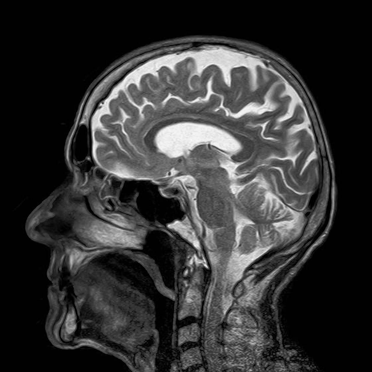
and my brain does something similar… In essence, my brain is simplifying the structure, highlighting some differences and reducing others. It’s filtering the image down to something like this:
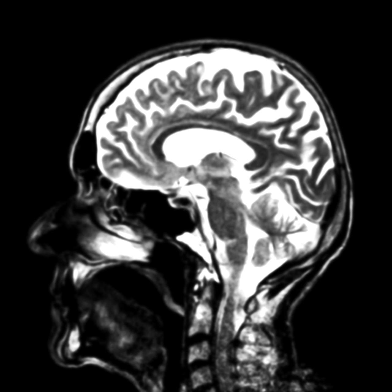
BUT the gradations of grey-scale on a sofa don’t mean the same thing as in an MRI scan of the brain. The original image actually contains far more gradations of grey than I can probably perceive…
But using Photoshop or another image processing program you can get the computer to mark them, and use false coloring to exaggerate the differences. Doing that to the original image produces this:
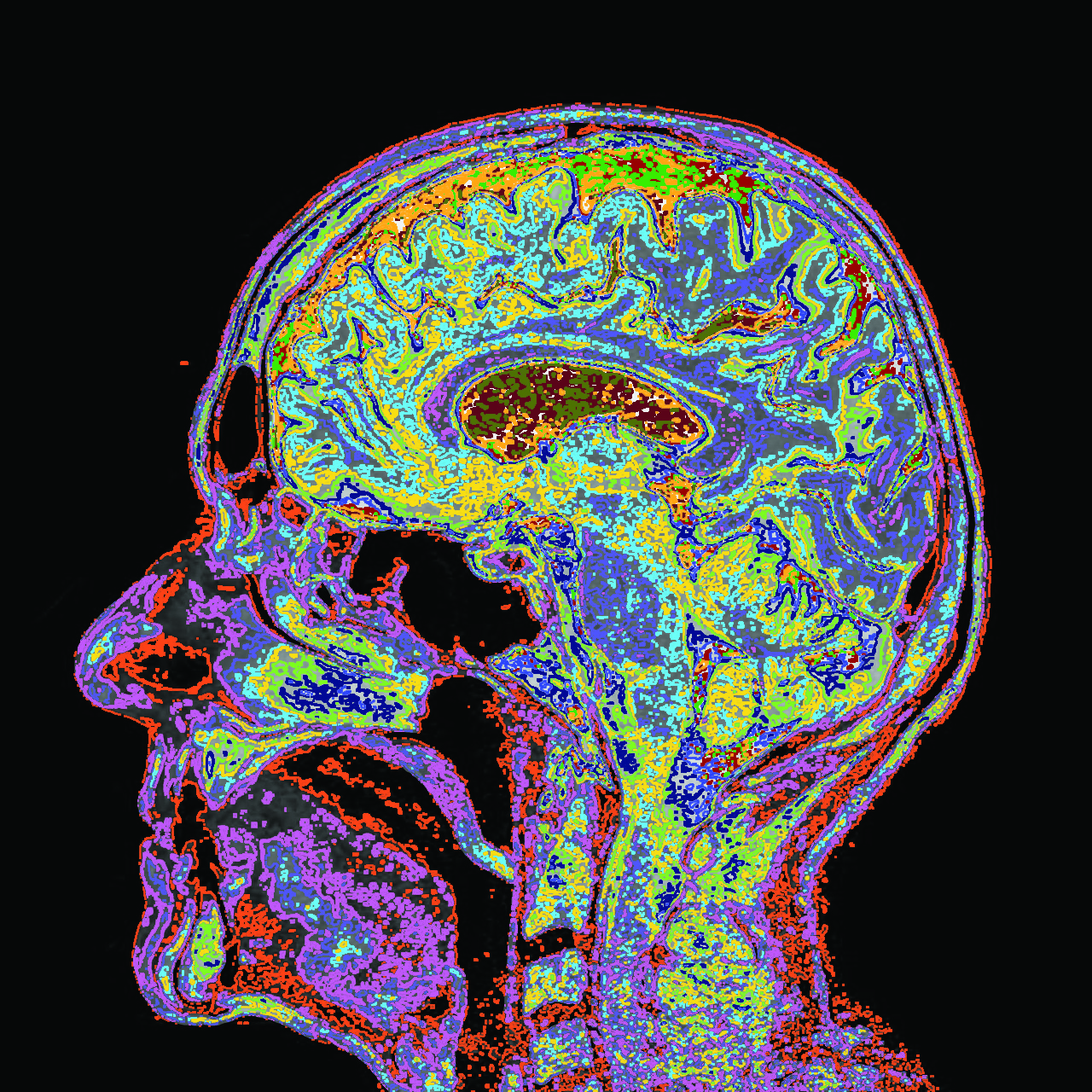
It’s not necessarily true that this rendering contains more functional information than the simpler one, but I’d bet it does. How meaningful are these new substructures? That’s for the experts to decide, but you have to notice them in the first place to ask the question.
The “ghosts” in this process are a level of visual processing that our brains often carry out below the surface, recognizing some shades of grey as the “same” and clustering them, ignoring others and filtering them out. There’s simply no guarantee that the way this is happening – trained by all kinds of situations in which we recognize patterns in images – will pick up the critical differences in an MRI image of the brain.
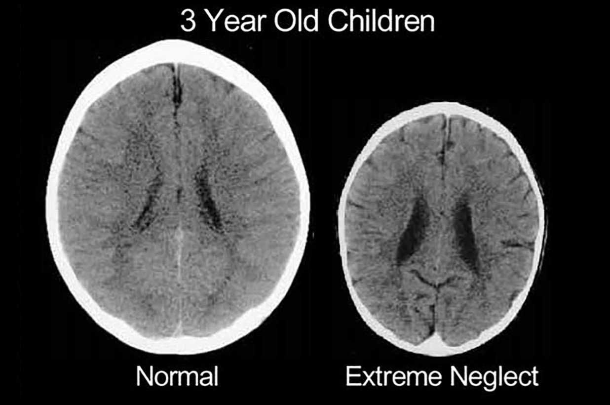
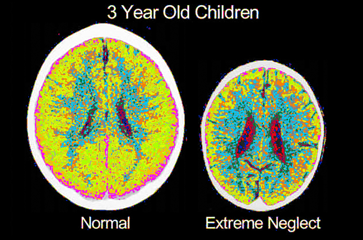
This morning I found a similar image in an article by the NY Post and used it to do the same thing. The piece refers to a study comparing the brains of a “normal, healthy” three-year-old and another who had suffered extreme emotional abuse. I’m not making any claims about the original study here, or the controls and so on, not having read it yet. Nor am I sure that the image they posted represents the original data, with the full resolution and color scale. But still, the difference is remarkable.
Here’s the image posted on the site:
And here’s my colorized version:
There’s certainly more to see. What does it mean? Thoughts are welcome.
